![]()
![]()
![]()
![]()
![]()
Image Generator to Support the Application of a Haptic Device for the Simulation of Arthroscopic Surgery
Functional Description
Name: Renata Zabawa
Advisor: Dr. Thomas L. Stewart
Date: October 31, 2005
Introduction:
Magnetic Resonance Imaging (MRI) uses the property of nuclear magnetic resonance to create images. An MRI creates hundreds of images that each shows a cross section of the knee. The cross sections of images can be stacked together to form a three dimensional image of the knee. The 3-D image is an alternative to analyzing the numerous separate cross sections. The goal of this project is to take the cross sections of a knee MRI and extract a 3-D model of the cartilage. This model is used to simulate a surgeon’s view during arthroscopic surgery. A second goal is to apply these results to simulate the arthroscopic surgery with a haptic feedback system. The simulation could be used as a training device for medical students.
Figure 1 shows a high-level block diagram for the entire system. A more detailed description of each subsystem follows.
DSP Software:
The input into the system is the MRI data points. Each scan is 500 X 500 pixels. The MRI scans have cross sections of the knee, which are taken and stacked together. DSP software uses Matlab to extract a three dimensional model of the cartilage from the MRI scans. The model of the cartilage shows the two menisci in the knee. Matlab is then used to separate the two menisci. This allows one menisci to be viewed at a time.
Graphics:
Once a 3-D image of the cartilage is created and the menisci are split, Matlab functions are used to put light on the cartilage and view it from different angles. These functions create a simulation of the surgeon’s view during arthroscopic surgery.
Display:
The simulation of an arthroscopic meniscus surgery is displayed on a computer monitor. Figure 2 and 3 exemplify the actual view a surgeon studies. Figure 2 illustrates torn menisci and Figure 3 shows healthy menisci.
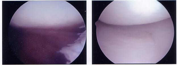
Figure 2: Arthroscopic Surgery View of Figure 3: Arthroscopic Surgery View of
Torn Cartilage Healthy Cartilage
References:
Links, Jonathan and Prince, Jerry. Medical Imaging: Signals and Systems. New Jersey: Pearson Education, 2006.