![]()
![]()
![]()
![]()
![]()
Image Generator to Support the Application of a Haptic Device for the Simulation of Arthroscopic Surgery
System Block Diagram
Name: Renata Zabawa
Advisor: Dr. Thomas L. Stewart
Date: November 4, 2005
Introduction:
The goal of this project is to take the cross sections of a knee MRI and extract a 3-D model of the cartilage. This model is used to simulate a surgeon’s view during arthroscopic surgery. A second goal is to apply these results to simulate the arthroscopic surgery with a haptic feedback system.
Block Diagrams:
Figure 1 shows the overall system block diagram. The input is the image data from the MRI scans. The MRI scans will be 500 X 500 pixels each. This data is used to create the model of the cartilage and generate the simulation of the arthroscopic surgery.
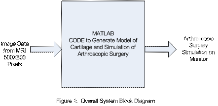
Figure 2 shows a high-level block diagram for the entire system. The MRI scan data points each show a cross sections of the knee. DSP software uses Matlab to generate a 3-D model of the cartilage. Then the model is used to create a simulation of an arthroscopic surgery.
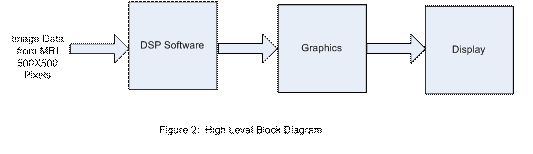
Figure 3 illustrates the DSP software used. Matlab loads the data from an MRI scan. To generate the three dimensional model of the cartilage, a few steps must be taken. First, the data is changed into the right format if it is needed. Then isosurfaces and isocaps are used on each set of MRI points. Isosurfaces use data that has been smoothed to display the overall structure of the knee. Isocaps use unsmoothed data to reveal details of the interior of the isosurfaces. The two surfaces created are used together to form a three dimensional model of the cartilage which shows the overall structure and the details of the interior of the knee. The model of the cartilage shows the two menisci in the knee. Matlab is then used to separate the two menisci allowing one meniscus to be viewed at a time.
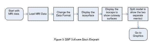
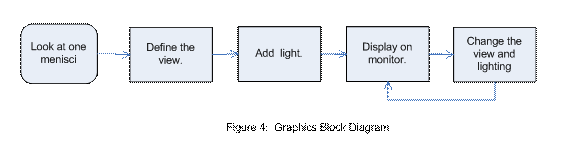
Figure 4 shows the graphics that will be accomplished with Matlab. Once a 3-D image of the cartilage is created and the menisci are split, Matlab functions are used to put light on the cartilage and view it from different angles. These functions create a simulation of the surgeon’s view during arthroscopic surgery. The simulation of an arthroscopic meniscus surgery is displayed on a computer monitor.