Speckle Reduction
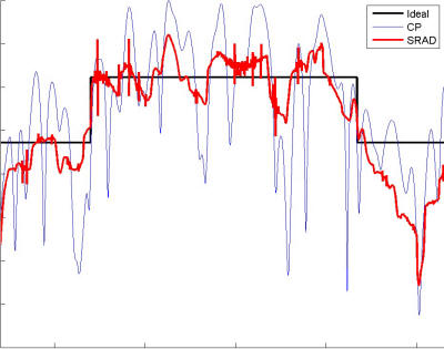
Below is an example of the effects of speckle on a target with a +6 dB contrast from the background. The black line shows an axial profile of the object being imaged. It has clearly defined edges as well as an obvious difference between the simulated lesion (raised region) and background. The blue shows how speckle corrupts the perceptibility of this object - regions in the background are at the same level as the target and regions in the target are at the same level as the background. After filtering the eREC-FC image with SRAD (red), two things happen: 1) the variance in the target region has decreased, and 2) a bias forms in the background region that pulls the levels down. This bias is the cause of the significant contrast improvement.
Below is a more extreme case of a +3 dB difference between target and background. Whereas in the +6 dB case above, a rough estimate could be made of lesion and background based on highest peak of the CP image, now no such estimate can be made. However, after eREC-FC and SRAD, the object is much more noticeable, thanks to the decreased variance in the target region and biases in the background.
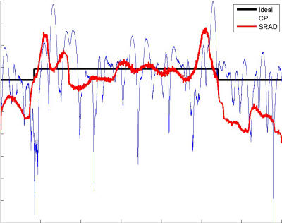
Photo Gallery
Speckle reduction